A Patient's Guide to Lumbar Disc Herniation
Introduction
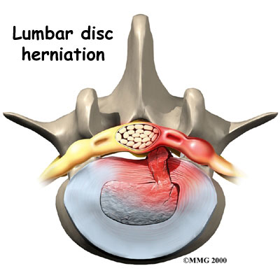
Although people often refer to a disc herniation as a "slipped disc," the disc doesn't actually slip out of place. Rather, the term "herniation" means that the material at the center of the disc has squeezed out of its normal space. This condition mainly affects people between thirty and forty years old.
This guide will help you understand
- how the problem develops
- how doctors diagnose the condition
- what treatment options are available
Chiropractic Treatment
First, surgery is the last option for disc problems in the lower back. In fact, 95% of patients with lower back pain with and without disc involvement do not require surgery. For these 95% of patients, conservative treatment is the recommended approach. Spinal misalignments, often painless at first, eventually lead to wear on the discs in the spine. The chiropractic approach to disc problems is to help restore better motion and position to the spinal joint. Besides reducing the disc bulging, better spinal function helps reduce inflammation and begin the slow process of healing the surrounding soft tissues. This is often combined with ice, traction, soft tissue massage, electrical muscle stimulation, ultrasound, traction, exercise and stretching. We do not always adjust or manipulate the spine in the presence of certain disc and nerve problems in the spine. Depending on your exam findings, other techniques can sometimes be used, although manipulation is usually very helpful.
The spine is made up of a series of connected bones called "vertebrae". The disc is a combination of strong connective tissues which hold one vertebra to the next, and acts as a cushion or shock absorber between the vertebrae. The disc is made of a tough outer layer called the "annulus fibrosis" and a gel-like center called the "nucleus pulposus." When healthy, discs allow normal turning and bending. As you get older, the center of the disc may start to lose water content, making the disc less effective as a cushion. This may cause a displacement of the disc's center (called a herniated or ruptured disc) through a crack in the outer layer. Most disc herniations occur in the bottom two discs of the lumbar spine, at and just below the waist.
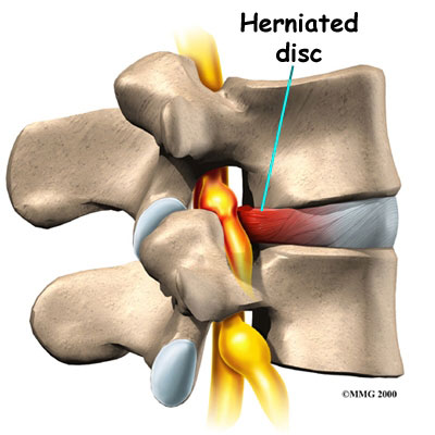
A herniated lumbar disc can press on the nerves in the spine and may cause pain, numbness, tingling or weakness of the leg. Similarly, a herniated or bulging disc in the neck may cause numbness, tingling or pain into the arm or hand. Herniated or bulging discs may also cause localized back or neck pain, although neck and back pain alone (without leg pain or arm pain) can have many causes other than a herniated disc.
Since spinal discs have a very poor blood supply, they depend upon the circulation of joint fluids to bring in nutrients and expel waste. If a spinal joint loses its normal motion and this pumping action is impaired, the health of the disc deteriorates. Like a wet sponge, a healthy disc is flexible. A dry sponge is hard, stiff, and can crack easily. This is how many disc problems begin.
Because of the way each disc is attached to the vertebra above and below it, a disc cannot "slip" as commonly thought. However, trauma or injury to the spine can cause discs to bulge, herniate, or worse, rupture. This can be quite painful, putting pressure on the spinal cord and nerve roots, interfering with their function. Disc problems are one of the most common conditions seen at the Winchester Hospital Chiropractic Center. Specially designed tables, called Flexion/Distraction tables are designed for treating such conditions.
Specific therapeutic exercises proven extremely effective in reducing disc problems in the lumbar spine will be demonstrated and performed in the office. These are called Mckenzie exercises or McKenzie Therapy. Traction can help de-compress the pressure on the discs of mid-back. Electrical muscle stimulation and ice are commonly used to help reduce the inflammation in the muscles and nerves. Longstanding poor posture is a common cause of disc problems in the low back. Therefore, postural corrective exercises will be prescribed. A thorough chiropractic examination and history will help in identifying the appropriate treatment for you. Chiropractic doctors see lumbar disc problems quite often in daily practice and have excellent results.
Anatomy
What parts of the spine are involved?
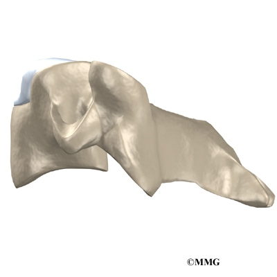
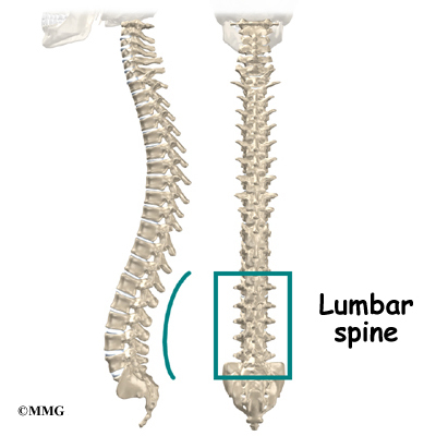
The human spine is formed by twenty-four spinal bones, called vertebrae. Vertebrae are stacked on top of one another to form the spinal column. The spinal column gives the body its form. It is the body's main upright support. The section of the spine in the lower back is known as the lumbar spine.
The lumbar spine is made of the lower five vertebrae. Doctors often refer to these vertebrae as L1 to L5. These five vertebrae line up to give the low back a slight inward curve. The lowest vertebra of the lumbar spine, L5, connects to the top of the sacrum, a triangular shaped bone at the base of the spine that fits between the two pelvic bones. Some people have an extra, or sixth, lumbar vertebra. This condition doesn't usually cause any particular problems.
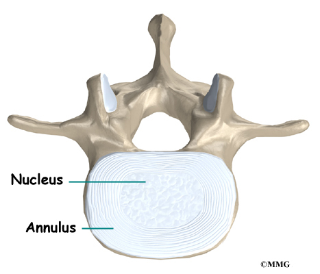
Intervertebral discs separate the vertebrae. The discs are made of connective tissue. Connective tissue is the material that holds the living cells of the body together. Most connective tissue is made of fibers of a material called collagen. These fibers help the disc withstand tension and pressure.
A disc is made of two parts. The center, called the nucleus, is spongy. It provides most of the disc's ability to absorb shock. The nucleus is held in place by the annulus, a series of strong ligament rings surrounding it. Ligaments are connective tissues that attach bones to other bones.
Healthy discs work like shock absorbers to cushion the spine. They protect the spine against the daily pull of gravity. They also protect it during strenuous activities that put strong force on the spine, such as jumping, running, and lifting.
Causes
Why do I have this problem?
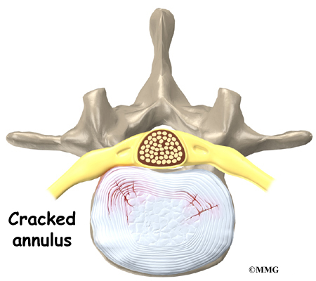
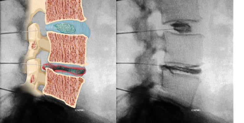
Herniation occurs when the nucleus in the center of the disc pushes out of its normal space. The nucleus presses against the annulus, causing the disc to bulge outward. Sometimes the nucleus herniates completely through the annulus and squeezes out of the disc.
Although daily activities may cause the nucleus to press against the annulus, the body is normally able to withstand this pressure. However, as the annulus ages, it tends to crack and tear. It is repaired with scar tissue. This process is known as degeneration. Over time, the annulus weakens, and the nucleus may begin to squeeze (herniate) through the damaged annulus. At first, the pressure bulges the annulus outward. Eventually, the nucleus may herniate completely through the outer ring of the disc.
Vigorous, repetitive bending, twisting, and lifting can place abnormal pressure on the shock-absorbing nucleus of the disc. If great enough, this increased pressure can injure the annulus, leading to herniation.
A lumbar disc can also become herniated during an acute, or sudden, injury. Lifting with the trunk bent forward and twisted can cause a disc herniation. A disc can also herniate from a heavy impact on the spine, such as falling from a ladder and landing in a sitting position.
Herniation causes pain from a variety of sources. It can cause mechanical pain. This is pain that comes from the parts of the spine that move during activity, such as the discs and ligaments. Pain from inflammation occurs when the nucleus squeezes through the annulus. The nucleus normally does not come in contact with the body's blood supply. However, a tear in the annulus puts the nucleus at risk for contacting this blood supply. When the nucleus herniates into the torn annulus, the nucleus and blood supply meet, causing a reaction of the chemicals inside the nucleus. This produces inflammation and pain. A disc herniation may also put pressure against a spinal nerve. Pressure on an irritated or damaged nerve can produce pain that radiates along the nerve. This is called neurogenic pain.
Symptoms
What does the condition feel like?
Many cases of lumbar disc herniation result from degenerative changes in the spine. The changes that eventually lead to a disc herniation produce symptoms gradually. At first, complaints may only include dull pain centered in the low back, pain that comes and goes over a period of a few years. Doctors think this is mainly from small tears in the annulus. Larger cracks in the annulus may spread pain into the buttocks or lower limbs.
When the disc herniates completely through the annulus, it generally causes immediate symptoms, with sharp pain that starts in one hip and shoots down part or all of the leg. Commonly, patients no longer feel their usual back pain, only leg pain. This is likely because painful tension on the annulus releases when the nucleus pushes completely through.
Disc herniations produce inflammation when the nucleus comes in contact with the body's blood supply (mentioned earlier). The inflammation can be a source of throbbing pain in the low back and may spread into one or both hips and buttocks.
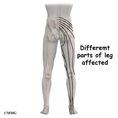
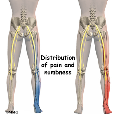
A herniated disc can press against a spinal nerve, producing symptoms of nerve compression. Nerve pain follows known patterns in the lower limbs. It can be felt on the side of the upper thigh, in the calf, or even in the foot and toes.
Pressure on the nerve can also cause sensations of pins, needles, and numbness where the nerve travels down the lower limbs. If this happens, a person's reflexes slow. The muscles controlled by the nerve weaken, and sensation in the skin where the nerve goes is impaired.
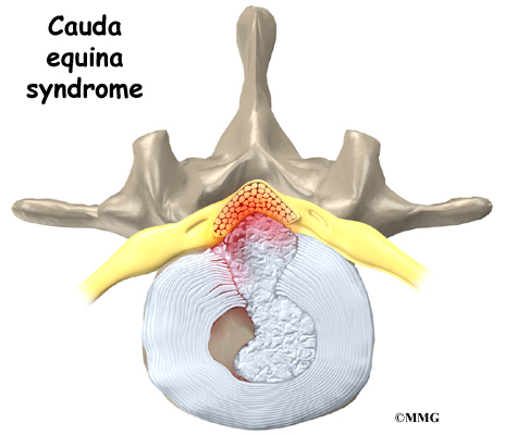
Rarely, symptoms involve changes in bowel and bladder function. A large disc herniation that pushes straight back into the spinal canal can put pressure on the nerves that go to the bowels and bladder. The pressure may cause low back pain, pain running down the back of both legs, and numbness or tingling between the legs in the area you would contact if you were seated on a saddle. The pressure on the nerves can cause a loss of control in the bowels or bladder. This is an emergency. If the pressure isn't relieved, it can lead to permanent paralysis of the bowels and bladder. This condition is called cauda equina syndrome. Doctors recommend immediate surgery to remove pressure from the nerves.
Diagnosis
How do doctors diagnose the problem?
Diagnosis begins with a complete history and physical exam. Your doctor will ask questions about your symptoms and how your problem is affecting your daily activities. These will include questions about where you feel pain and whether you have numbness or weakness in your legs. Your doctor will also want to know what positions or activities make your symptoms worse or better. Doctors rely on your report of pain to get an idea which disc is causing problems and if a nerve is being squeezed.
Then the doctor examines you to determine which back movements cause pain or other symptoms. Your skin sensation, muscle strength, and reflexes are also tested.
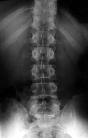
X-rays are of minor help in diagnosing disc herniations. The discs don't actually show up on X-rays. However, doctors can tell if the space between the vertebrae is smaller than normal. This can be an indication that wear and tear on one or more discs is causing problems. However, many peoples' X-rays show degeneration of the discs. This is because degeneration in the discs is part of aging, like skin that wrinkles with time.
Computed tomography (a CT scan) may be ordered. This is a detailed X-ray that lets doctors see "slices" of the body's tissue. The image can show if a herniated disc is putting pressure on a spinal nerve.
Doctors may combine the CT scan with myelography. To do this, a special dye is injected into the space around the spinal canal--the subarachnoid space. When the CT scan is performed, the dye highlights the spinal cord and nerves. The dye can improve the accuracy of a standard CT scan for diagnosing a herniated disc.
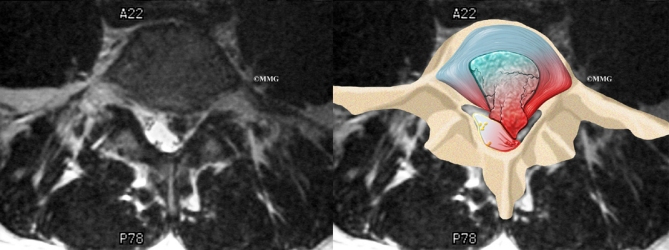
When more information is needed, your doctor may order magnetic resonance imaging (MRI). The MRI machine uses magnetic waves rather than X-rays to show the soft tissues of the body. It gives a clear picture of the discs and whether a herniation is present. Like the CT scan, this machine creates pictures that look like slices of the area your doctor is interested in. The test does not require special dye or a needle.
Doctors sometimes order a specialized X-ray test called discography. In this test, dye is injected into one or more discs. The dye is seen on X-ray and can give some information about the health of one or more discs. This test may be used when surgery is being considered to determine which disc is causing problems.
Doctors may also order electrical tests to locate more precisely which spinal nerve is being squeezed. Several tests are available to see how well the nerves are functioning, including the electromyography (EMG) test. This test measures how long it takes a muscle to work once a nerve signals it to move. The time it takes will be slower if a herniated disc has put pressure on a spinal nerve. Another test is the somatosensory evoked potential (SSEP) test. The SSEP is used to measure nerve sensations. These sensory impulses travel up the nerve, informing the body about sensations such as pain, temperature, and touch. The function of a nerve is recorded by an electrode placed over the skin in the area where the nerve travels. Doctors will often run these tests before performing surgery for a lumbar disc herniation.
Other Treatment Options
Surgery
If the symptoms you feel are mild and there is no danger they'll get worse, surgery is not usually recommended. However, if signs appear that pressure is building on the spinal nerves, surgery may be required--sometimes right away. The signs doctors watch for when reaching this decision include weakening in the leg muscles, pain that won't ease up, and problems with the bowels or bladder.
Surgical treatment for lumbar disc herniation includes
- laminectomy and discectomy
- microdiscectomy
- posterior lumbar fusion
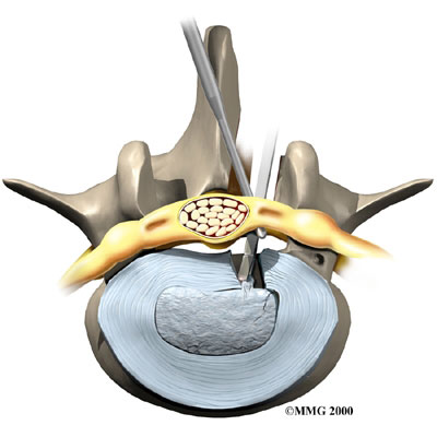
Laminectomy and Discectomy
The lamina forms a roof-like structure over the back of the spinal canal. In this procedure, a thumbnail-sized piece of the lamina is removed (laminectomy) so the surgeon can more easily take out the problem disc (discectomy). This procedure is mainly used when the herniated disc is putting pressure on a nerve and causing pain to spread down one leg.
 Microdiscectomy
Microdiscectomy is becoming the standard surgery for lumbar disc herniation. The procedure is used when a herniated disc is putting pressure on a nerve root. It involves carefully taking out part of the problem disc (discectomy). By performing the operation with a surgical microscope, the surgeon only needs to make a very small incision in the low back. Categorized as "minimally invasive surgery," this surgery is thought to be less taxing on patients. Advocates also believe that this type of surgery is easier to perform, that it prevents scarring around the nerves and joints, and that it helps patients recover more quickly.
Posterior Lumbar Fusion
Lumbar disc herniation causes mechanical pain, the type of pain caused by wear and tear in the parts of the lumbar spine. Fusion surgery is mainly used to stop movement of the painful area by joining two or more vertebrae into one solid bone. This keeps the bones and joints from moving, easing mechanical pain.
In this procedure, the surgeon lays small grafts of bone over the problem area on the back of the spinal column. Most surgeons will also apply metal plates and screws to prevent the problem vertebrae from moving. This protects the graft so it can heal better and faster.
All content provided by eORTHOPOD® is a registered trademark of Medical Multimedia Group, L.L.C.. Content is the sole property of Medical Multimedia Group, L.L.C.. and used herein by permission.
All materials from eORTHOPOD® are the sole property of Medical Multimedia Group, L.L.C.. and are used herein by permission. eORTHOPOD® is a registered trademark of Medical Multimedia Group, L.L.C..
|