A Patient's Guide to Lumbar Spinal Stenosis
Introduction
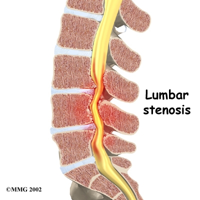
Stenosis means "closing in." Spinal stenosis describes a condition in which the nerves in the spinal canal are "closed in," or compressed. The spinal canal is the hollow tube formed by the bones of the spinal column. Anything that causes this bony tube to shrink can squeeze the nerves inside. As a result of many years of wear and tear on the parts of the spine, the tissues nearest the spinal canal sometimes press against the nerves. This helps explain why lumbar spinal stenosis (stenosis of the low back) is a common cause of back problems in adults over fifty-five years old.
This guide will help you understand
- how the problem develops
- how doctors diagnose the condition
- what treatment options are available
Chiropractic Treatment
Manual treatments such as spinal manipulation and lumbar spinal traction have been shown to be effective in reducing the symptoms associated with lumbar spinal stenosis. Patients often find that curling up to sleep or lying on their back with their knees bent and supported gives them relief. These positions flex the spine forward, which widens the spinal canal and can give the spinal cord some more room to breath. You will be shown specific spinal postures aimed at decreasing pain, improving mobility, strength and function. Patients are taught how to protect their spines. Treatments are often combined with electrical muscle stimulation, ice, ultrasound, soft tissue massage, and traction. All of these are designed to help decrease pain and inflammation and improve function and mobility. Please see treatment sections to read further about these procedures.
Another technique performed only by chiropractic doctors is called manual flexion/distraction which is an advanced and specific form of traction. It involves the use of a specially designed traction table which can help open and widen the spinal canal, taking pressure off of the spinal cord. Patients often feel immediate relief after the first treatment! On rare occasions, bracing may be indicated to help keep the spine in a semi-flexed position, widening the spinal canal.
Aerobic exercises may include walking on a treadmill, riding a stationary bike, or swimming. Stationary biking is a good exercise because it keeps the spine slightly bent forward, opening the spinal canal. These activities can also relieve the stress of low back pain, improve heart and lung health, endurance and can cause your body to release endorphins into the blood stream. Endorphins are the body's own natural painkillers.
When needed, patients are encouraged to take certain actions to improve their spine health. Patients who smoke are encouraged to get help to quit. Because of the limited blood supply in the tissues of the low back, smoking speeds the degenerative process and impairs healing. Patients who are out of shape are encouraged to get fit. This strategy makes it less likely that back pain or injury will strike again in the future.
Dr. Kane and Dr. Zohn will show you how to keep your spine safe during routine activities. You'll learn about healthy posture and how posture relates to the future health of your spine. You'll learn about body mechanics, how the body moves and functions during activity. Chiropractors teach safe body mechanics to help you protect the low back as you go about your day. This includes the use of safe positions and movements while lifting and carrying, standing and walking, and performing work duties.
Anatomy
What part of the back is involved?
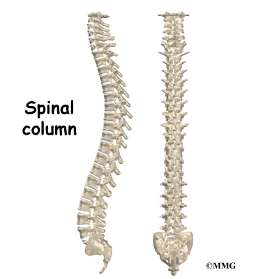
The human spine is made up of twenty-four spinal bones, called vertebrae. Vertebrae are stacked on top of one another to create the spinal column. The spinal column gives the body its form. It is the body's main upright support.
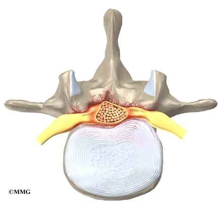
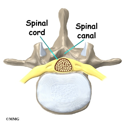
The back portion of the spinal column forms a bony ring. When the vertebrae are stacked on top of each other, these bony rings create a hollow tube. This bony tube, called the spinal canal, surrounds the spinal cord as it passes through the spine. Just as the skull protects the brain, the bones of the spinal column protect the spinal cord.
The spinal cord only extends to the second lumbar (low back) vertebra. Below this level, the spinal canal encloses a bundle of nerves that go to the lower limbs and pelvic organs. The Latin term for this bundle of nerves is cauda equina, meaning "horse's tail."
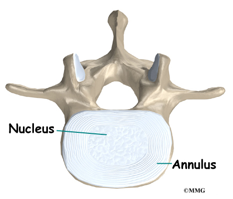
An intervertebral disc fits between each vertebral body and provides a space between the spine bones. The disc normally works like a shock absorber. It protects the spine against the daily pull of gravity. It also protects the spine during heavy activities that put strong force on the spine, such as jumping, running, and lifting.
An intervertebral disc is made up of two parts. The center, called the nucleus, is spongy. It provides most of the ability to absorb shock. The nucleus is held in place by the annulus, a series of strong ligament rings surrounding it. Ligaments are strong connective tissues that attach bones to other bones.
Causes
Why do I have this problem?
In the lumbar spine, the spinal canal usually has more than enough room for the spinal nerves. The canal is normally seventeen to eighteen millimeters around, slightly less than the size of a penny. Spinal stenosis develops when the canal shrinks to twelve millimeters or less. When the size drops below ten millimeters, severe symptoms of lumbar spinal stenosis occur.
There are many reasons why symptoms of spinal stenosis develop. Some of the more common reasons include
- congenital stenosis (being born with a small spinal canal)
- spinal degeneration
- spinal instability
- disc herniation
Congenital stenosis: Some people are born with a spinal canal that is narrower than normal. They may not feel problems early in life. However, having a narrow spinal canal puts them at risk for stenosis. Even a minor back injury can cause pressure against the spinal cord. People born with a narrow spinal canal often have problems later in life, because the canal tends to become narrower due to the effects of aging.
Degeneration: Degeneration is the most common cause of spinal stenosis. Wear and tear on the spine from aging and from repeated stresses and strains can cause many problems in the lumbar spine. The intervertebral disc can begin to collapse and the space between each vertebrae shrinks. Bone spurs may form that stick into the spinal canal and reduce the space available to the spinal nerves. The ligaments that hold the vertebrae together may thicken and also push into the spinal canal. All of these conditions cause the spinal canal to narrow.
View animation of degeneration
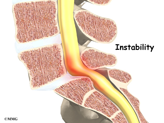
Spinal instability: Spinal instability can cause spinal stenosis. Spinal instability means that the bones of the spine move more than they should. Instability in the lumbar spine can develop if the supporting ligaments have been stretched or torn from a severe back injury. People with diseases that loosen their connective tissues may also have spinal instability. Whatever the cause, extra movement in the bones of the spine can lead to spinal stenosis.
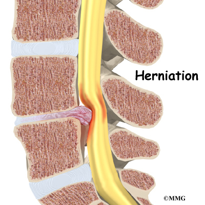
Disc herniation: Spinal stenosis can occur when an intervertebral disc in the low back herniates. Normally, the shock-absorbing disc is able to handle the downward pressure of gravity and the strain from daily activities. However, if the pressure on the disc is too strong, such as landing from a fall in a sitting position, the nucleus inside the disc may rupture through the outer annulus and squeeze out of the disc. This is called a disc herniation. If an intervertebral disc herniates straight backward, it can press against the nerves in the spinal canal, causing symptoms of spinal stenosis.
Symptoms
What does the spinal stenosis feel like?
Spinal stenosis usually develops slowly over a long period of time. This is because the main cause of spinal stenosis is spinal degeneration in later life. Symptoms rarely develop quickly when degeneration is the source of the problem. A severe injury or a herniated disc may cause symptoms to develop immediately.
Patients with stenosis don't always feel back pain. Primarily, they have pain and weakness in their legs--usually in both legs at the same time. Some people say they feel that their legs are going to give out on them.
Symptoms mainly affect sensation in the lower limbs. Nerve pressure from stenosis can cause a feeling of pins and needles in the skin where the spinal nerves travel. Reflexes become slowed. Some patients report "charleyhorses" in their leg muscles. Others report strange sensations like water trickling down their legs.
Symptoms change with the position of the low back. Bending forward (flexion) widens the spinal canal and usually eases symptoms. That's why people with stenosis tend to get relief when they sit down or curl up to sleep. Activities such as reaching up, standing, and walking require the spine to straighten or even bend back slightly (extension). This position of the low back makes the spinal canal smaller and often worsens symptoms.
Diagnosis
How do doctors diagnose the problem?
Diagnosis begins with a complete history and physical examination. Your doctor will ask questions about your symptoms and how your problem is affecting your daily activities. This will include questions about your pain or if you have feelings of numbness or weakness in your legs. Your doctor will also want to know whether your symptoms are worse when you're up standing or walking and if they go away when you sit down.
The doctor does a physical examination to see which back movements cause pain or other symptoms. Your skin sensation, muscle strength, and reflexes are also tested.
X-rays can show if the problems are from changes in the bones of the spine. The images can show if degeneration has caused the space between the vertebrae to collapse. X-rays may also show any bone spurs sticking into the spinal canal.
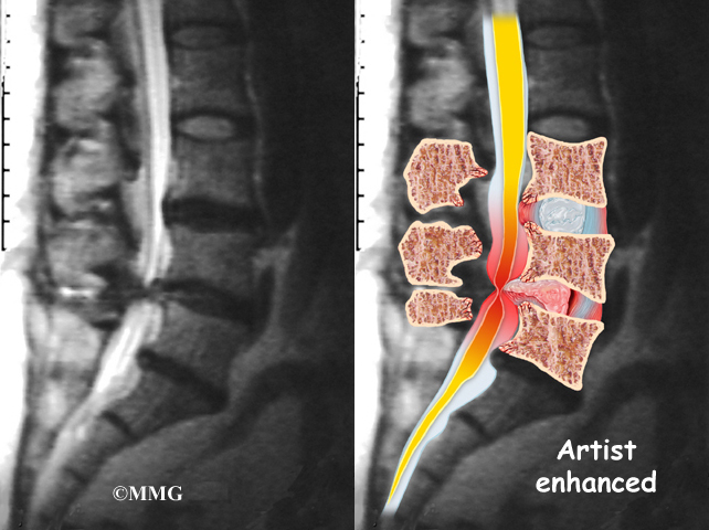
When more information is needed, your doctor may order an MRI scan (magnetic resonance imaging scan). The MRI machine uses magnetic waves rather than X-rays to show the soft tissues of the body. This test gives a clear picture of the spinal canal and whether the nerves inside are being squeezed. This machine creates pictures that look like slices of the area your doctor is interested in. The test does not require dye or a needle.
Computed tomography (a CT scan) may be ordered. The CT scan is a detailed X-ray that lets your doctor see "slices" of bone tissue. The image can show any bone spurs that may be sticking into the spinal column and taking up space around the spinal nerves.
When the diagnosis is still not clear, doctors may recommend electrical tests of the nerves that go to the legs and feet. An electromyogram (EMG) checks whether the "motor" pathway of a nerve is working correctly. Motor impulses travel down the nerve and work to energize muscles.
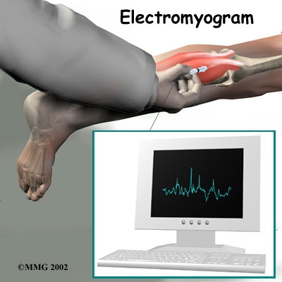
Doctors may also order a somatosensory evoked potential (SSEP) test to locate more precisely where the spinal nerves are being squeezed. The SSEP is used to measure nerve sensations. These sensory impulses travel up the nerve, informing the body about sensations such as pain, temperature, and touch. The function of a nerve is recorded by an electrode placed over the skin in the area where the nerve travels.
Not all causes of spinal stenosis are from degenerative conditions. Doctors use blood tests to determine whether symptoms are coming from other conditions, such as arthritis or infection.
All content provided by eORTHOPOD® is a registered trademark of Medical Multimedia Group, L.L.C.. Content is the sole property of Medical Multimedia Group, L.L.C.. and used herein by permission.
All materials from eORTHOPOD® are the sole property of Medical Multimedia Group, L.L.C.. and are used herein by permission. eORTHOPOD® is a registered trademark of Medical Multimedia Group, L.L.C..
|