A Patient's Guide to Osteoarthritis of the Knee

Introduction
Osteoarthritis (OA) is a common problem for many people after middle age. OA is sometimes referred to as degenerative, or wear and tear, arthritis. OA commonly affects the knee joint. In fact, knee OA is the most common cause of disability in the United States. In the past, people were led to believe that nothing could be done for their problem. Now doctors have many ways to treat knee OA so patients have less pain, better movement, and enhanced quality of life.
This guide will help you understand
- how OA develops
- how OA of the knee causes problems
- how doctors treat the condition
Chiropractic Treatment
Chiropractic treatment plays a critical role in the treatment of knee OA. A primary goal is to help you learn how to control symptoms and maximize the health of your knee. You will learn ways to calm pain and symptoms, which might include the use of rest, heat, or topical rubs.
Chiropractic adjustments/manipulations are performed to help restore normal biomechanics to the knee joint. This is where chiropractic manipulation has an advantage over other specialties. It is important to manipulate and stretch capsular joint restrictions during the treatment phase. This helps improve normal motion and keeps the joint lubricated. Modalities such as ice, electrical muscle stimulation, ultrasound and heat may also be used to decrease pain and inflammation.
Range-of-motion and stretching exercises will be used to improve knee motion. You will be shown strengthening exercises for the hip and knee to help steady the knee and give additional joint protection from shock and stress. People with knee OA who have strong leg muscles enjoy fewer symptoms and prolong the life of their knee joint. Your therapist will also suggest tips for getting your tasks done with less strain on the joint.
You will learn ways to calm pain and symptoms, which might include the use of rest, heat, ice or topical rubs. Range-of-motion and stretching exercises will be used to improve hip motion. You will be shown strengthening exercises for the hip to steady the joint and protect it from shock and stress. We can also suggest tips for getting your tasks done with less strain on the joint.
Many Practitioners frequently overlook the feet when examining the hip. The feet should always be checked for poor or faulty biomechanics. The feet are the foundation of the body and many foot conditions eventually contribute to knee, hip and lower back problems. Orthotics for your shoes and sneakers will have a significant effect in reducing excessive biomechanical forces on the hip and the shock-absorbing component will reduce impact and protect the joint. People who walk regularly are encouraged to choose a soft walking surface, such as a cinder or grass track. At the Winchester Hospital Chiropractic Center, we can take molds of your feet and fit you for custom made orthotics.
OA can't be cured, but therapies are available to ease symptoms and to slow down the degeneration. Recent information shows that mild cases of knee OA may be maintained and in some cases improved without surgery.
Medical studies have shown that glucosamine and chondroitin sulfate can also help people with OA. These supplements seem to have nearly the same benefits as anti-inflammatory medicine with fewer side affects. Many doctors feel the research supports these supplements and are encouraging their patients to use them.
Dr. Zohn and Dr. Kane will teach their patients how to protect the arthritic knee joint. This starts with tips on choosing activities that minimize impact and twisting forces on the knee. People who modify their activities can actually slow down the affects of knee OA. For instance, people who normally jog might decide to walk, bike, or swim to reduce impact on their knee joint. Sports that require jumping and quick starts and stops may need to be altered or discontinued to protect the knee joint.
A new type of knee brace, called a knee unloading brace, can help when OA is affecting one side of the knee joint. For example, a bowlegged posture changes the way the knee joint lines up. The inside (medial) part of the knee joint gets pressed together. The cartilage suffers more damage, and greater pain and problems occur. The unloading brace pushes against the outer (lateral) surface of the knee, causing the medial side of the joint to open up. In this way, the brace shares the pressure and unloads the arthritic medial side of the joint. A knee unloading brace can help relieve pain and allow people to do more of their usual activities.
For some cases of knee OA, you may be given a heel wedge to wear in your shoe. By tilting the heel, the wedge alters the way the knee lines up, which works like the unloading brace mentioned above to take pressure off the arthritic part of the knee.
Anatomy
Which parts of the knee are affected?
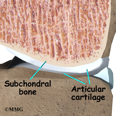
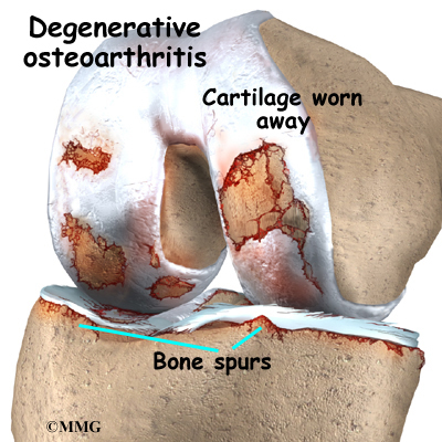
The main problem in OA is degeneration of the articular cartilage. Articular cartilage is the smooth lining that covers the ends of the leg bones where they meet to form the knee joint. The cartilage gives the joint freedom of movement by decreasing friction. The layer of bone just below the articular cartilage is called subchondral bone.
When the articular cartilage degenerates, or wears away, the bone underneath is uncovered and rubs against bone. Small outgrowths called bone spurs or osteophytes may form in the joint.
Causes
How does knee OA develop?
OA of the knee can be caused by a knee injury earlier in life. It can also come from years of repeated strain on the knee. Fractures of the joint surfaces, ligament tears, and meniscal injuries can all cause abnormal movement and alignment, leading to wear and tear on the joint surfaces. Not all cases of knee OA are related to a prior injury, however. Scientists believe genetics makes some people prone to developing degenerative arthritis. Obesity is linked to knee OA, and losing only ten pounds can reduce the risk of future knee OA by 50 percent.
Scientists believe that problems in the subchondral bone may trigger changes in the articular cartilage. Normally, the articular cartilage protects the subchondral bone. But some medical conditions can make the subchondral bone too hard or too soft, changing how the cartilage normally cushions and absorbs shock in the joint.
Symptoms
What does knee OA feel like?
OA develops slowly over several years. The symptoms are mainly pain, swelling, and stiffening of the knee. Pain is usually worse after activity, such as walking. Early in the course of the disease, you may notice that your knee does fairly well while walking, then after sitting for several minutes your knee becomes stiff and painful. As the condition progresses, pain can interfere with simple daily activities. In the late stages, the pain can be continuous and even affect sleep patterns.
Diagnosis
How do doctors identify OA?
The diagnosis of OA can usually be made on the basis of the initial history and examination.
X-rays can help in the diagnosis and may be the only special test required in the majority of cases. X-rays can also help doctors rule out other problems, since knee pain from OA may be confused with other common causes of knee pain, such as a torn meniscus or kneecap problems. In some cases of early OA, X-rays may not show the expected changes.
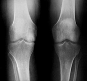
Magnetic resonance imaging (MRI) may be ordered to look at the knee more closely. An MRI scan is a special radiological test that uses magnetic waves to create pictures that look like slices of the knee. The MRI scan shows the bones, ligaments, articular cartilage, and menisci. The MRI scan is painless and requires no needles or dye.
If the diagnosis is still unclear, arthroscopy may be necessary to actually look inside the knee and see if the joint surfaces are beginning to show wear and tear. Arthroscopy is a surgical procedure in which a small fiber-optic TV camera is inserted into the knee joint through a very small incision, about one-quarter of an inch long. The surgeon can move the camera around inside the joint while watching the pictures on a TV screen. The structures inside the joint can be poked and pulled with small surgical instruments to see if there is any damage.
All content provided by eORTHOPOD® is a registered trademark of Medical Multimedia Group, L.L.C.. Content is the sole property of Medical Multimedia Group, L.L.C.. and used herein by permission.
All materials from eORTHOPOD® are the sole property of Medical Multimedia Group, L.L.C.. and are used herein by permission. eORTHOPOD® is a registered trademark of Medical Multimedia Group, L.L.C..
|